


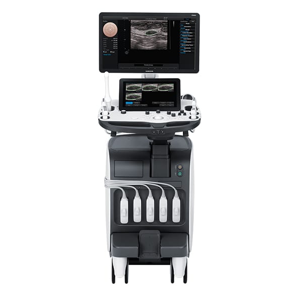
Samsung RS 80A
High-resolution images for confident diagnoses
Enhanced technologies that expand capabilities
The advanced technical capabilities that the RS80A with Prestige features are built on the successes of Samsung technologies, including superior image quality, while offering exclusive options. The features such as S-Fusion and S-Shearwave provide diagnostic confidence and user convenience in challenging practices.
 S-Vision Beamformer
 S-Vision Beamformer
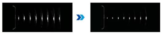
The S-Vision beamformer demonstrates a clearer image that receives returning signals through a sophisticated digital filtering system resulting in reduced side lobes, less noise and artifact. It enhances the image quality with better clarity and consistent results.
Essential tools for interventional procedures
Samsung pushes the boundaries of ultrasound technology. With leading technologies like S-Fusion and S-Tracking, you can expect accuracy in interventional procedures./h3>
With advanced technologies like S-Shearwave and CEUS+thenumber of biopsies can be reduced, lesions become visible and examinations are easier to perform.
S-Fusion
 S-Fusion
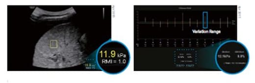
S-Shearwave detects the velocity of the shearwave propagated through the targeted lesion and displays the numerical measurement of stiffness in kPa or m/s together with a Reliable Measurement Index (RMI)*. Also it provides Variation Range (VR), a range of value, that intuitively shows the uniformity of tissue stiffness in the Region of Interest (ROI). The wider range means the less tissue stiffness uniformity. In the profile window, the user can easily edit each measurement value depending on its Reliable Measurement Index. S-Shearewave helps to reduce the number of conventional liver biopsies by providing quantitative tissue characteristic information.
CEUS+
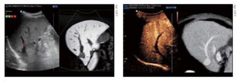 CEUS+
CEUS+CEUS+ technology uses the unique properties of ultrasound contrast agents. When excited with a Low MI the oscillating micro bubbles reflect both the basic frequencies and harmonic signals. In the CEUS+ harmonic image on the left the perfused parts are displayed and on the right side a conventional B-Mode image.
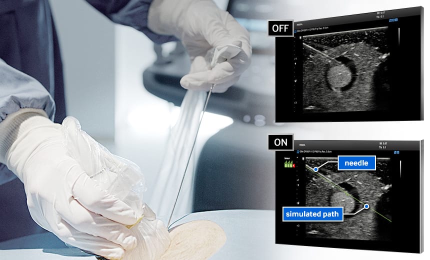
S-Tracking
S-Tracking increases the rate of accuracy during interventional procedures by providing the simulated path of the needle and the target mark in the live ultrasound image. Clear Track, one of two functions provided by S-Tracking, secures the accuracy by using a specialized needle with a sensor tip. Virtual Track uses general needles during the procedure, providing both accuracy and economic benefit.
Advances that minimize risk
Early detection of cardiovascular diseases and risk for stroke
Advanced QuickScan™
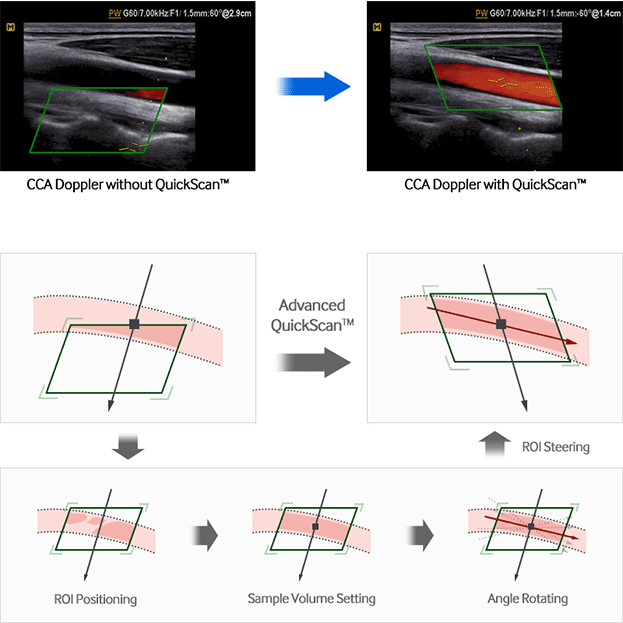
Advanced QuickScanTM technology provides intuitive optimization of gray scale and Doppler parameters. One touch of the QuickScanTM button elevates efficiency and workflow by adjusting functions including color gain and color box location.
Auto IMT+™
Auto IMT+™ is a screening tool that analyzes a patient’s risk of strokeand heart disease. It allows easy intima-media thickness measurement of both the anterior and posterior wall of the common carotid by clicking a button. This simple procedure enhances exam productivity and adds diagnostic value.
Arterial Analysis
Arterial Analysis detects functional changes of vessels, providing measurement values such as the stiffness and intima-mediathickness. Since the functional changes occur before morphological changes, this technology supports the early detection of cardiovascular diseases..
Strain+
Strain quantitatively displays a Bull’s Eye which shows left ventricular motion and dyssynchrony at a glance.
Stress Echo
The Stress Echo package includes wall motion scoring and reporting. It includes exercise Stress Echo, pharmacologic Stress Echo, diastolic Stress Echo and free programmable Stress Echo.
Standardized analysis and classification
For a better ultrasound breast assessment Samsung offers a wide range of useful imaging and quantitative tools.
S-Detect™ for breas
S-Detect™ employs Breast Imaging-Reporting and Data System (BI-RADS®) scores for standardized analysis and classification of suspicious lesions. By simply clicking the suspected area, it draws the lesion area and provides the characteristics of the lesion and a recommendation on whether the lesion is benign or malignant. Such technology assists in a more accurate diagnosis, while improving the efficiency of workflow and reducing the time users spend in repetitive tasks.
E-Breast™ (ElastoScan™ for breast)
 E-Breast™ (ElastoScan™ for breast)
E-Breast™ is a technology that calculates the strain ratio between the selected target and surrounding fatty tissues. Unlike conventional ultrasound elastography, E-Breast™ requires only one ROI to be selected by the user. This simplified process enhances consistency and reduces the chance of error by eliminating the step of manual selection of the surrounding fatty tissue region..
3D Volume linear transducer
Multi-dimensional volume data acquired by Volume linear transducer visualizes the structure of targeted planes in a single step. It helps users to deliver more accurate and efficient diagnosis..
Superior image quality with enhanced technology
Departments


Urology

Ophthalmology

Bone Density

Endocrinology

Ultrasound

Cardiology

Χειρουργικής Θυρεοειδούς και Ενδοκρινών Αδένων

Cytology
