


Contact Us
Email: info@medicus-center.com
Phone: 210 890 0909

DIGITAL RADIOLOGY DEPARTMENT FOR DEMANDING IMAGING REQUIREMENTS


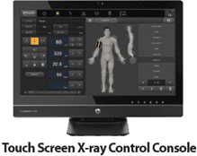
Our diagnostic center is equipped with the state-of-the-art GXR-40SD digital radiography system by DRGEM, which features:
Automatic exposure system Rotating lamp
Reduced radiation dose Wide range of exposure
Convenience in performing scans and patient-friendly
X-rays of high digital quality
Radiography is to this day the most common type of imaging in medicine. During this technique, dynamic images are taken in real time, depicting the morphology of the human body. The most common radiology scans include:
Chest X-ray
Sinus X-ray
Spine X-ray
Head X-ray
Hip X-ray
Abdominal X-ray
Bone age X-ray
The key benefits of the modern GXR-40SD digital radiography system are:
Very low radiation dose / Maximum radiation protection.
Excellent image quality / Better diagnosis.
High image quality even in large magnification, when focusing on a specific area of interest.
Elimination of technical errors associated with X-ray views and the need for frequent repeat exams.
Minimum scan time.
Difficult to diagnose cases, as it can depict with extreme resolution microfractures and orthopedic cases that cannot be easily diagnosed (e.g. scaphoid area).
Available to doctors at all times, for reading and comparative evaluation. 100% synchonization between doctor and radiologist.
Suitable for young patients due to the low radiation dose, but also ideal even for people who cannot move easily and need something convenient and quick.
It offers fast and convenient access to diagnostic data for referring/attending physicians, even remotely with safe internet connection.
The radiology center can store the scans in an organized manner, through the comprehensive picture and communication system (PACS), for up to 10 years.
The images are stored in digital format and remain unchanged.
Lower cost (film printing is optional).
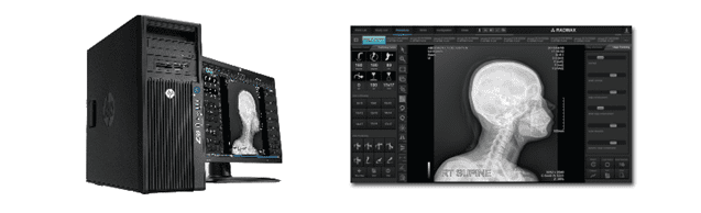
The state-of-the-art DRGEM GXR-40SD digital radiography system is equipped with a modern-design, latest-technology DRGEM Detector, which offers all the benefits of digital technology, such as easy to use, excellent image quality and great configuration capabilities, including:
Contrast and brightness adjustment in an area of interest.
Contrast and resolution improvement.
Reduced image noise / rejection of artifacts.
Option of printing multiple views on a single film.
Measurement of distances, angles, Cobb angle.
Actual printing size.
Impressive reduction of radiation dose, even by 40%.
It takes just a few seconds and, depending on the area the radiography is performed, patients may be standing, seated or lying down.
High image quality and ability to focus on a specific area of interest.
The image is instantly transmitted to the DRGEM console in DICOM format, where it is analyzed, processed and then sent to our diagnostic center’s central picture and communication system (PACS).
The procedure is very simple and quick.
The X-rays are available on radiology film, CD and USB or may be sent via email to the attending physician (Dicom Server).
Digital radiography is one of the most significant advancements, as it reduces both the exposure time and the radiation dose compared to conventional X-rays on film or Computer Radiology (CR) used by most radiology center these days, whereby the analog film is rendered in digital format.
Digital radiography has less radiation than analog and does not require as many repetitions.
The most significant factor is the exposure time.
In conventional radiography, 4-5 sec. of exposure to radiation are required to get a single X-ray.
Digital radiography reduces the exposure time to 0.25-0.04 sec., with images of higher resolution and contrast, less radiation (2-3 times lower dose) and less time to capture the image.
The main advantage of digital radiography is that the doctor can process the image and make a diagnosis, which they either may have avoided doing with analog X-rays or may have required an additional X-ray to view the problem.
The projections are performed by a qualified radiology technician, who ensures the highest possible diagnostic value for every diagnostic problem.
