


Telephone: +30 210 890 0909
Email: info@medicus-center.com
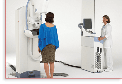
The laboratory is equipped with a Senographe Essential General Electric high-tech digital mammography device.
The mammography reports are issued immediately on the spot, without making the patients wait or call them back.
The top quality of our equipment and the experience of the medical staff make stereotactic breast biopsies and hook installation a short and painless procedure.

ANALOG – DIGITAL MAMMOGRAPHY AND THEIR ROLE IN BREAST CANCER DIAGNOSIS
Mammography is an X-ray method that uses low-dose X-rays to examine the breast.
The aim of mammography is the early diagnosis of breast cancer by detecting small tumors and microcalcifications.
Mammography has significantly reduced breast cancer deaths (30-35%).
Mammography is the only radiology method that, combined with self-examination and clinical examination by qualified doctors, contributes significantly to the early diagnosis of breast cancer.
In medically advanced countries, all women over the age of 40 undergo annual mammography screening.

Mammography is clearly the method of choice for breast screening and may be supplemented, where appropriate, by breast ultrasound, galactography and breast MRI.
Breast ultrasound is considered necessary for women younger than 40 years of age, and is particularly useful for characterizing masses depicted in a mammography, as well as for investigating palpable masses not depicted in a mammography.
The rate of false negative results in mammography ranges from 6 to 10%.
The fact that a small percentage of breast cancers escape the attention of radiology specialists is usually due to increased breast density, but also to the indistinguishable differences of cancer compared to the normal mass gland (breast gland).
Further screening is required in 7-10% of women undergoing preventive mammography screening. When a woman is recalled by a doctor for additional testing, this can be particularly stressful. Of the women recalled for additional testing, 10% undergo a breast biopsy and 3.5% are diagnosed with breast cancer.

The exposure of the breast to radiation during a mammogram is a problem for a significant proportion of the female population.
Long-term studies show that, for each death of a woman over 40 years of age attributable to radiation, there are 48.5 women rescued from breast cancer.
The radiation dose to which a woman is exposed during a mammogram is equal to the cosmic radiation she receives in a 3-hour flight.
The effort of medical technology to improve the quality and accuracy of mammography resulted in a revolutionary application, that of digital mammography.
Digital mammography uses X-rays to produce breast images.
However, digital mammography does not use films but a special X-ray detector, which converts radiation into light.
The light is then converted into a digital signal received on a computer screen.
In the US, only 8% of diagnostic units are equipped with a digital mammograph.
Large-scale studies carried out over the last decade in North America and Europe have demonstrated the superiority of digital mammography in the following groups of women:
Women under 50
Women of all ages with very dense breasts
Premenopausal or perimenopausal women of all ages

The cost of digital mammography is currently a major disadvantage, as a digital mammography device is 4-5 times more expensive than a conventional one. In contrast to this digital mammography disadvantage, experts contrast a number of advantages:
Short examination time
It takes 1-2 minutes for digital mammography while the corresponding time for a conventional mammogram is 15 minutes.
Automatic enlargement of breast areas without additional irradiation.
Less time required for biopsies or breast needle localization.
The radiological image is edited to achieve optimum quality.
Use of diagnostic aids such as computer-assisted diagnosis.
All of the patient’s mammograms are stored in an electronic file.
Use of telemedicine to enable and facilitate the second opinion of a radiology specialist.
3D imaging with relatively low radiation doses.
The first major medical study demonstrating the contribution of preventive mammography to rescuing large numbers of women from breast cancer was published in 1967.
In the decades that followed, there have been some small as well as large advancements in the field of medical technology, such as digital mammography.
However, regardless of the state of the art, the war against breast cancer will only be won by realizing the value of preventive screening.